In advanced material characterisation, understanding both how a surface looks and what it is made of is essential. A material may appear smooth and uniform under a microscope, but could contain impurities or hidden compositional variations that affect performance. This is where the combined power of SEM + EDS becomes invaluable.
Scanning Electron Microscopy (SEM) reveals surface morphology, while Energy Dispersive Spectroscopy (EDS) identifies elemental composition. Together, they provide a complete picture of structure and chemistry at the micro-scale.
What Is SEM?
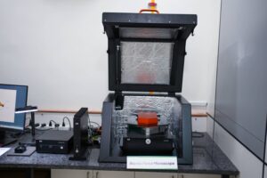
SEM (Scanning Electron Microscopy) is a technique used to examine surface structure at very high magnifications. It works by generating an electron beam from a tungsten filament and focusing it onto the sample using condenser and objective lenses.
When the focused electron beam strikes the surface, signals are produced. These signals are converted into detailed grayscale images showing:
- Surface texture
- Particle size and distribution
- Micro-cracks and defects
- Coating layers
- Morphological features
SEM images are typically captured at resolutions such as 30 µm, 10 µm, and 5 µm, depending on magnification needs.
What Is EDS?
EDS (Energy Dispersive Spectroscopy) is an analytical technique attached to the SEM system. While SEM focuses on structure, EDS determines elemental composition.
When the electron beam hits the sample, it not only produces imaging signals but also causes atoms in the material to emit characteristic X-rays. Each element emits X-rays at specific energy levels. The EDS detector measures these energies and converts them into a spectrum.
The result is:
- A graph showing energy peaks
- Identification of elements present
- Estimation of percentage composition
How SEM + EDS Work Together
SEM and EDS operate simultaneously within the same chamber. After placing the mounted sample on the stage, the chamber is sealed to create vacuum conditions.
Once imaging begins:
- 1. The electron beam scans the surface.
- 2. SEM detectors produce morphological images.
- 3. EDS detectors collect characteristic X-rays.
- 4. Software generates both image and elemental data.
Standard output typically includes 8 SEM images and 2 EDS spectra per sample, with optional elemental mapping reports.
Analysis time:
- SEM only: ~45 minutes
- SEM + EDS: ~60 minutes
Difference Between SEM Imaging and EDS Mapping
Although both use the same electron beam, their outputs are fundamentally different.
1. SEM Image (Morphology)
An SEM image is a grayscale micrograph that shows:
- Shape
- Surface roughness
- Cracks or fractures
- Particle clusters
- Surface coatings
It is purely structural information. You can see features clearly, but you cannot confirm what those features are chemically made of.
For example:
You might see bright and dark regions in an SEM image. Without EDS, you cannot determine whether those regions represent different elements or just surface topography differences.
2. EDS Spectrum (Element Identification)
The EDS spectrum appears as a graph with peaks corresponding to different elements.
- The horizontal axis shows energy.
- The vertical axis shows intensity.
- Each peak represents a specific element.
For example:
A peak at a particular energy level might confirm the presence of titanium (Ti), zinc (Zn), silver (Ag), or other elements.
EDS provides:
- Qualitative identification (which elements exist)
- Semi-quantitative estimation (approximate percentage)
3. Elemental Composition Mapping
Elemental mapping is an advanced EDS feature where the system generates a colour-coded distribution map of elements across the scanned surface.
Instead of just listing elements, mapping shows:
- Where each element is located
- Whether elements are uniformly distributed
- Presence of contamination or segregation
In mapping:
- One colour may represent iron.
- Another colour may represent oxygen.
- A third colour may represent carbon.
This allows researchers to visually confirm whether elements are evenly dispersed or clustered.
Why SEM + EDS Is Important
Using SEM alone provides only half the story. Using EDS alone provides composition but no structural context.
Combined, SEM + EDS allows you to:
- Confirm material purity
- Detect contamination
- Verify alloy composition
- Study corrosion or oxidation
- Analyse coatings
- Investigate failure mechanisms
For example:
If a surface crack is observed under SEM, EDS can determine whether corrosion products are present inside the crack.
Sample Preparation Requirements
Since SEM operates under a vacuum, only dried samples are allowed. Non-conducting samples require gold coating to prevent charging.
Proper mounting on standard discs (50 mm or 125 mm) ensures stable imaging and accurate analysis.
Incorrect preparation may lead to distorted images or inaccurate elemental readings.
Real-World Applications
SEM + EDS is widely used in:
- Metal alloy verification
- Nanomaterial research
- Battery materials
- Pharmaceutical analysis
- Polymer characterisation
- Surface coating validation
It helps industries ensure product quality and reliability before large-scale manufacturing.
Final Comparison Summary
| Feature | SEM | EDS |
|---|---|---|
| Purpose | Surface structure | Elemental composition |
| Output | Grayscale images | Energy spectrum + mapping |
| Shows shape? | Yes | No |
| Shows elements? | No | Yes |
| Mapping available? | No | Yes |
Conclusion
SEM + EDS combines visual imaging with chemical identification. SEM shows the “face” of the material: its texture, cracks, and particles. EDS reveals the “ingredients”: the elements that make up those structures.
Together, they provide a powerful, integrated method for surface and compositional analysis. By understanding the difference between SEM images and elemental mapping, students and researchers can better interpret results and make informed material decisions.