The morning had been theoretical: two speakers, an hour each, on the science of microbial bioactives and engineered biofactories. The afternoon was where that science met the centre’s instruments. Five research-cadre faculty ran three rotating stations until every student had operated everything.
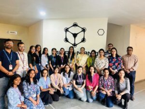
On 11 April 2026, after the two morning lecture sessions of the MNRDC one-day workshop on microbial cells as green bio-foundries at Parul University, the seventeen enrolled students were divided into three groups. From 1:30 PM to 4:00 PM, the groups rotated through three hands-on stations operated by the research cadre of the Micro-Nano Research and Development Center. The workshop kit each student received at the 10:00 AM registration included a brochure with the full equipment catalogue of the MNRDC. The afternoon was the working demonstration of that catalogue
The workshop kit and the brochure that listed the day's instruments
Registration started at 10:00 AM. Each student received a workshop kit containing a folder with a diary, a pen, the day’s schedule, and the MNRDC equipment brochure.
The brochure was not promotional material. It was a working catalogue of the instruments and facilities at the Micro-Nano Research and Development Center (MNRDC) at Parul University. For many of the seventeen students, this was the first time they were reading a complete inventory of what the centre actually holds, and for several of them, this was the first time they would later operate those instruments under direct faculty supervision. The brochure mapped the workshop’s afternoon directly. Each station was selected from instruments documented in the catalogue.
The MNRDC suite of published articles covers each instrument category in detail. Students who completed the workshop and wanted reference material to return to could read the Scanning Electron Microscopy (SEM-EDS) facility article, the X-Ray Diffraction (XRD) services article, the Atomic Force Microscopy (AFM) capability article, the consolidated material characterisation lab walkthrough, and the top material tests for PhD research across mechanical, materials, and chemical engineering, each of which documents the equipment used in the workshop’s afternoon stations in production-research context.
The two coordinators: Dr. Juhi Saxena and Dr. Anwesha Khanra
Two faculty members coordinated the workshop at the operational level. Both were responsible for managing the day’s activities and leading specific sessions within the overall programme.
Dr. Juhi Saxena, a faculty member in the Faculty of Applied Sciences at Parul University, is recognised among the institution’s seven Stanford-Elsevier Global Top 2% Scientists. She introduced the opening session delivered by Dr. Anupam Jyoti and guided the academic flow of the workshop from beginning to end. Her research profile reflects the university’s strength in applied sciences and supports the institution’s ability to host expert sessions such as those delivered by Dr. Gunjan Sharma of Gujarat Biotechnology University.
Dr. Anwesha Khanra served as the second workshop coordinator and also supervised the microalgae cultivation station during the afternoon practical session. Her dual role connected the morning’s discussions on algal biofactories with hands-on laboratory work, allowing participants to observe the practical aspects of algal cultivation. Algae as solar-powered, carbon-neutral biofactories are discussed in the Dr. Gunjan Sharma session deep-dive article, while the workshop provided participants with the opportunity to see these concepts demonstrated in practice.
The five research-cadre faculty who ran the afternoon
The afternoon practical sessions were conducted by five research faculty members who guided participants through different laboratory demonstrations and research facilities.
- Dr. Mahendra Singh Rathore: Led the sputtering demonstration, introducing participants to a sample preparation and surface modification technique widely used in thin-film research and high-resolution microscopy. The session explained both the underlying principles and the operation of the sputtering equipment available at the MNRDC.
- Dr. Vishal Mehta: Conducted the MNRDC facility walkthrough, explaining how the centre’s research instruments work together within an integrated workflow. Participants were introduced to the laboratory layout, equipment arrangement, and the sequence through which research samples move during characterisation.
- Dr. Meenu Khan: Led the scanning electron microscopy (SEM) micrography session using microbial cell samples. The demonstration highlighted sample preparation methods, imaging resolution, and the use of SEM for studying microbial morphology.
- Dr. Anwesha Khanra: Served as both workshop coordinator and faculty lead for the microalgae cultivation laboratory. Her station demonstrated live microalgae cultures, cultivation conditions, and the laboratory setup supporting algal biofactory research, reinforcing concepts introduced during the morning sessions.
- Dr. Mohit Tannarana: Conducted demonstrations on UV-FTIR spectroscopy and Atomic Force Microscopy (AFM), introducing participants to analytical techniques used for molecular characterisation and surface topology analysis in biological and materials research.
MNRDC Bio-Foundry Workshop – The Science Behind SCFAs, Gut Health & Chronic Disease
Station 1: SEM micrography on microbial cells, led by Dr. Meenu Khan
Scanning electron microscopy (SEM) is a widely used imaging technique for high-resolution surface characterisation of microbial cells. During the workshop, Dr. Meenu Khan’s demonstration enabled participants to observe cellular structures that had been discussed during the morning’s biological sessions.
The session introduced the fundamentals of SEM, including sample preparation procedures such as dehydration, specimen mounting, and sputter coating with a conductive material. Participants also learned the basic principles of electron beam imaging, the resolution capabilities of SEM, and the types of microbial surface features that can be examined using this technique.
The practical demonstration closely complemented the morning’s lectures. Images of neutrophil extracellular traps (NETs) presented by Dr. Anupam Jyoti had been generated using scanning electron microscopy, allowing students to directly relate the laboratory equipment to the research examples discussed earlier. Similarly, SEM is one of the analytical techniques used in studies examining exopolysaccharide (EPS) characterisation.
The MNRDC’s broader scanning electron microscopy capabilities are described in the dedicated SEM-EDS facility article, which outlines both the centre’s research services for external users and its role in practical student training.
Station 2: Microalgae cultivation, led by Dr. Anwesha Khanra
The algae laboratory serves as the MNRDC’s working bio-foundry, where participants were introduced to practical aspects of microalgae cultivation. Dr. Anwesha Khanra’s station translated the morning’s discussion of algae as solar-powered, carbon-neutral biofactories into a hands-on laboratory experience.
The session covered live microalgae cultivation, including the factors that influence growth such as light intensity, temperature, nutrient composition, salinity, and aeration. Participants also learned about the organisation of the algae laboratory as an active research facility and the considerations involved in scaling laboratory cultivation for industrial and commercial applications.
Potential applications discussed during the session included biofuel production, omega-3 fatty acid extraction, beta-carotene production, and the development of agricultural biostimulants. These examples demonstrated how microalgae research extends beyond laboratory studies into a range of biotechnology and sustainability applications.
The practical session closely complemented Dr. Gunjan Sharma’s earlier presentation. Microalgal species discussed during the morning lecture—including Chlorella vulgaris, Haematococcus pluvialis, Dunaliella salina, Nannochloropsis, Schizochytrium, Crypthecodinium cohnii, and Porphyridium purpureum—were presented within the context of operational cultivation systems, helping participants connect theoretical concepts with laboratory practice.
Station 3: Combined MNRDC walkthrough with UV-FTIR, AFM, and sputtering
The third station provided a comprehensive introduction to multiple research techniques through a combination of a facility walkthrough led by Dr. Vishal Mehta, a UV-FTIR and Atomic Force Microscopy (AFM) demonstration by Dr. Mohit Tannarana, and a sputtering demonstration by Dr. Mahendra Singh Rathore. Together, these sessions illustrated how different analytical techniques contribute to a complete research workflow.
- MNRDC facility walkthrough | Dr. Vishal Mehta: The walkthrough introduced participants to the layout of the MNRDC laboratories, the placement of major research instruments, and the sequence through which samples move during characterisation. It also explained how students interact with research-active faculty and laboratory facilities, providing the broader context for the demonstrations that followed.
- UV-FTIR and Atomic Force Microscopy (AFM) | Dr. Mohit Tannarana: The session introduced ultraviolet Fourier-transform infrared (UV-FTIR) spectroscopy as a technique for molecular characterisation. Participants learned how FTIR spectroscopy is used to analyse compounds such as extracted exopolysaccharides, complementing topics discussed during the morning lectures. The demonstration also covered Atomic Force Microscopy (AFM), which measures nanoscale surface topography by mapping height variations, providing information that complements the surface images produced by scanning electron microscopy (SEM).
- Sputtering | Dr. Mahendra Singh Rathore: The sputtering demonstration explained how a thin conductive coating, typically gold or platinum, is deposited onto biological samples before SEM imaging. This preparation step prevents electron charging during microscopy and is an essential part of the SEM workflow. The session helped participants understand how sample preparation directly influences the quality of electron microscopy images.
The dedicated AFM capability article provides additional information about the MNRDC’s Atomic Force Microscopy facility, including its applications in surface roughness analysis, nano-imaging, and external research services.
Why the three-station rotation matters: connecting morning theory to afternoon instrumentation
The structure of the afternoon practical sessions was intentionally designed so that each laboratory station reinforced concepts introduced during the morning lectures. By rotating through every station, participants were able to connect theoretical knowledge with the research instruments and techniques used to generate scientific evidence.
- Dr. Anupam Jyoti’s morning session | Cytoprotection, dysbiosis, bacteriocins, and exopolysaccharides: The scanning electron microscopy (SEM) images of neutrophil extracellular traps (NETs) presented during the lecture were produced using the same imaging technique demonstrated in the afternoon. Similarly, the exopolysaccharide (EPS) characterisation studies discussed during the session employ Fourier-transform infrared (FTIR) spectroscopy, allowing participants to observe both analytical methods in practice.
- Dr. Gunjan Sharma’s morning session | Bacterial, fungal, yeast, algal, and plant biofactories: The discussion of algal biofactories was reinforced through the live microalgae cultivation station, where participants observed operational culture systems. Topics related to biosynthetic gene clusters and metabolic engineering were further complemented by demonstrations of molecular and surface characterisation techniques presented at the combined instrumentation station.
This integrated approach strengthened the learning experience by linking scientific concepts with laboratory practice. Participants who studied neutrophil extracellular trap (NET) formation during the morning sessions were later able to observe the microscopy techniques used to generate such images. Likewise, discussions of microalgal biofactories were reinforced through direct exposure to live cultivation systems, helping connect theoretical knowledge with real research environments.
The valedictory workflow: Q&A, certificate distribution, and the Google feedback form
At 4:00 PM, after the three-station rotation was completed, the workshop reconvened for the valedictory session.
- Q&A session: Direct interaction with the faculty cadre on specific instrument questions, technique selection for hypothetical research questions, and follow-up on the morning’s content.
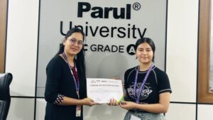
- Certificate distribution: Each of the seventeen enrolled students received a workshop completion certificate, providing formal documentation of participation that students could attach to their academic record and future research applications.
- Google feedback form: A structured written-response form circulated for completion. The form captured both quantitative ratings and free-text written responses on what had worked and what could be improved. The workshop coordinators’ assessment, drawn from the returned forms, was that the day had landed well across the enrolled cohort.
How the MNRDC research cadre connects to Parul University's broader research infrastructure
The five research-cadre faculty who ran the workshop in the afternoon are not exclusively workshop staff. They are research-active faculty whose work feeds into Parul University’s broader research output. The institution holds NAAC A++ accreditation with a CGPA of 3.55. Government-funded research currently stands at more than Rs 58.31 crore across 315 funded projects. Seven faculty members are in the Stanford-Elsevier global top 2 per cent of scientists, including Dr. Juhi Saxena. The Faculty of Applied Sciences, Parul Institute of Applied Sciences (PIAS), and the MNRDC together form the working research stack into which workshops like the April 2026 session on microbial cells as green bio-foundries are embedded.
The broader research and innovation ecosystem at Parul University extends beyond applied sciences. The Parul Innovation and Entrepreneurship Research Centre (PIERC) supports student startups across the institution, including portfolio cases that have raised external investment. The institution’s coordinated research-and-development infrastructure means that workshops at the MNRDC are connected to an ecosystem of research, innovation, and entrepreneurship that students can access through multiple pathways.
FAQs
Who are the MNRDC research cadre faculty?
The MNRDC research cadre comprises five research-active faculty members at Parul University's Micro-Nano Research and Development Center. Dr. Mahendra Singh Rathore leads sputtering and sample-preparation work. Dr. Vishal Mehta leads facility walkthroughs and research workflow coordination. Dr. Meenu Khan leads scanning electron microscopy (SEM) work, including specific applications on microbial cell samples. Dr. Anwesha Khanra (also a workshop coordinator) leads microalgae cultivation in the algae lab. Dr. Mohit Tannarana leads UV-FTIR (ultraviolet Fourier-transform infrared) spectroscopy and atomic force microscopy (AFM) demonstrations. Together, this cadre ran the afternoon hands-on stations at the 11 April 2026 workshop on microbial cells as green bio-foundries.
Who coordinated the MNRDC workshop on microbial bio-foundries?
The workshop on Microbial Cells as Green Bio-Foundries for Therapeutics and Environmental Remediation, held at the MNRDC on 11 April 2026, was coordinated by Dr. Juhi Saxena and Dr. Anwesha Khanra. Dr. Juhi Saxena is listed among Parul University's seven Stanford-Elsevier global top 2 per cent scientists. She introduced the morning session by Dr. Anupam Jyoti and managed the academic framing of the workshop. Dr. Anwesha Khanra served as the second coordinator and also led the afternoon microalgae cultivation station in the algae lab.
What instruments did the seventeen workshop participants use in the afternoon?
The seventeen enrolled students were divided into three groups that rotated through three hands-on stations from 1:30 PM to 4:00 PM until every student had operated every instrument. Station 1 was scanning electron microscopy (SEM) on microbial cell samples, led by Dr. Meenu Khan. Station 2 was microalgae cultivation in the algae lab, led by Dr. Anwesha Khanra. Station 3 was a combined facility walkthrough led by Dr. Vishal Mehta, a UV-FTIR and atomic force microscopy (AFM) demonstration led by Dr. Mohit Tannarana, and a sputtering introduction led by Dr. Mahendra Singh Rathore. The three-station structure ensured that every student had hands-on exposure to five different instrument categories in a single afternoon.
How does the afternoon hands-on session connect to the morning lectures?
The afternoon stations were selected to operationalise the morning's content directly. Dr. Anupam Jyoti's session on microbial bioactives included neutrophil extracellular trap (NET) imagery that comes from scanning electron microscopy and exopolysaccharide characterisation studies that use FTIR spectroscopy. Both techniques were operationally available at the afternoon stations. Dr. Gunjan Sharma's session on engineered biofactories included algal biofactories (Chlorella, Haematococcus, Nannochloropsis), which became operational rather than theoretical at the microalgae cultivation station. The connection between morning theory and afternoon instrumentation was structural rather than illustrative, allowing students to learn one thing across two layers
What is the MNRDC equipment brochure and what does it cover?
The MNRDC equipment brochure, distributed to each of the seventeen enrolled students in the workshop kit at 10:00 AM registration, is the working catalogue of instruments and facilities at the Micro-Nano Research and Development Center at Parul University. The brochure documents scanning electron microscopy (SEM), X-ray diffraction (XRD), atomic force microscopy (AFM), UV-FTIR spectroscopy, sputtering, microalgae cultivation, and related capabilities. Each instrument category is also documented in dedicated articles on the Parul University blog, including the SEM-EDS facility article, the XRD services article, the AFM capability article, the consolidated material characterisation lab walkthrough, and the top material tests for PhD research article. The brochure mapped the workshop's afternoon stations directly, with each station selected from instruments documented in the catalogue.
Can students from outside the Faculty of Applied Sciences access MNRDC workshops?
MNRDC workshops are open to students of Parul University's Faculty of Applied Sciences and the Parul Institute of Applied Sciences (PIAS), as well as students from related faculties whose academic interests align with the workshop topic. The 11 April 2026 workshop on Microbial Cells as Green Bio-Foundries was attended by seventeen students with backgrounds appropriate to microbiology, biotechnology, and applied sciences. Beyond workshops, the MNRDC operates as a research service centre for external clients and as a teaching facility for student users across the institution. The centre's working catalogue documents the service offering for both student users and external research clients.
What is the connection between MNRDC workshops and Parul University's broader research record?
The MNRDC workshop programme is one node in a wider research ecosystem at Parul University. The institution holds NAAC A++ accreditation with a CGPA of 3.55. Government-funded research currently stands at more than Rs 58.31 crore across 315 funded projects. Seven faculty members are in the Stanford-Elsevier global top 2 per cent of scientists, including Dr. Juhi Saxena, who coordinated this workshop. The Inflammation Research Lab at the Parul Institute of Applied Sciences, led by Dr. Anupam Jyoti, the speakers' programme that brought Dr. Gunjan Sharma from Gujarat Biotechnology University, and the broader Faculty of Applied Sciences together form the working research stack into which MNRDC workshops are embedded. The infrastructure tested in a one-day workshop is the same infrastructure that supports research-cadre projects and funded research throughout the year.