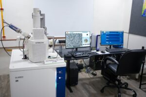
The Bruker D6 PHASER at MNRDC has analysed over 600 samples using standard XRD, which identifies crystal structure and material phases through Bragg’s Law diffraction. Standard XRD is covered in detail in the Centre’s dedicated XRD explainer article. This article focuses on what happens beyond standard XRD: the four advanced analytical modes available on the same instrument that address research questions standard XRD cannot answer.
Read more on – XRD Explained: Bragg’s Law, Peaks, and Material Identification
XRR: X-Ray Reflectivity for Thin Film Analysis
XRR is the specialised technique that complements standard XRD. While XRD examines the internal crystal structure of a material (the atomic skeleton), XRR examines the layered structure of thin films deposited on substrates. The distinction is important: XRD tells what the material is made of; XRR tells how thick, how smooth, and how dense a coating is.
How and what XRR Measures
- Thickness – Layers from 1 nanometre to 500 nanometres approximately.
- Roughness – the smoothness or bumpy surface at the microscopic scale.
- Density – packed material within layers.
How to Read an XRR Graph
Unlike the sharp spikes of a standard XRD diffractogram, an XRR graph produces a series of fading oscillatory waves. The wave pattern carries specific information about the film:
- Wide waves: the layer is very thin
- Narrow/tight waves: the layer is thick
- Fast fading: the surface is rough
- Slow fading: the surface is smooth (mirror-like)
At MNRDC, XRR is available as an add-on to standard XRD analysis. Users must request XRR specifically when submitting samples. This is particularly relevant for researchers working on thin film coatings deposited using the MNRDC’s own HHV Auto 500 sputtering system, because the deposit-then-characterise workflow happens entirely within the same facility.
Thin Film Coating for Solar Cells & Semiconductors at MNRDC, Parul University!
GIXRD: Grazing Incidence X-Ray Diffraction for Surface Coatings
Standard XRD sends the X-ray beam through the full depth of a sample. When analysing a thin coating on a thick substrate, the beam passes through the coating entirely and hits the substrate beneath. The resulting diffractogram shows 99% substrate signal and almost no coating signal. The coating data is effectively invisible.
GIXRD solves this by changing the geometry. Instead of hitting the sample from above at varying angles, the beam strikes the surface at a tiny, fixed grazing angle, typically less than 1 degree. At this shallow angle, the X-rays penetrate only the topmost layer of the material and never reach the substrate. The diffraction data comes exclusively from the coating.
Read more on – SEM Testing at MNRDC
When Researchers Should Request GIXRD Instead of Standard XRD
- Scratch-resistant coatings on glass (Gorilla Glass-type surface analysis)
- TiN hard coatings on tools deposited by sputtering
- ITO transparent electrodes on solar cell substrates
- Anti-reflective coatings on optical components
- Any thin film where the coating thickness is under 500 nm and the substrate would dominate standard XRD
The analogy: standard XRD is like an X-ray of bones through skin. GIXRD is like examining only the tattoo on the skin surface without seeing the bones at all.
Read more about – XRD vs SEM vs AFM: Which Test for Your Sample
TOPAS: Rietveld Refinement for Quantitative Phase Analysis
TOPAS (Total Pattern Analysis Solutions) is the advanced analysis software that transforms XRD from qualitative identification (what phases are present) into quantitative analysis (exactly how much of each phase is present, with mathematical certainty).
Read more about – MNRDC Facilities and Services
How Rietveld Refinement Works
Standard XRD peak matching tells a researcher that their sample contains Phase A and Phase B. Rietveld refinement tells them the sample is 73.2% Phase A and 26.8% Phase B, with calculated uncertainty margins. This process is divided into 3 major steps –
Step 1 – The researcher tells TOPAS about phases expectation in samples. (Example – 80% Cellulose and 20% Epoxy)
Step 2 – TOPAS initiates the calculation of the theoretical diffraction pattern that should come out on the basis of crystallographic physics for that particular composition.
Step 3 – TOPAS mindfully compares the pattern (calculated) against the real measured pattern and adjusts thousands of parameters until both patterns match.
Step 4 – The entire output is a list of quantities: phase fractions, refined crystal parameters, and a goodness-of-fit metric that proves the analysis is valid. It is the standard for publication-quality quantitative XRD in journals.
Real-World Example: Process Optimisation
XRD combined with TOPAS functions as a before-and-after fitness comparison for materials. A steel component is scanned before heat treatment; the graph shows a soft Austenite phase. After quenching in oil, the graph shows peaks shifted to hard Martensite. If the after-treatment graph still shows residual Austenite, the engineer knows the furnace temperature needs to increase. TOPAS quantifies exactly how much unconverted Austenite remains, giving the engineer a precise target for process adjustment.
Read more about – Top Material Tests for PhD Research
ICDD PDF-4: The Global Reference Library for Material Identification
The International Centre for Diffraction Data (ICDD) maintains the world’s most comprehensive database of diffraction patterns. MNRDC uses PDF-4, the most advanced paid version of this database, which contains over one million reference patterns. Each material has a unique Powder Diffraction File (PDF) that specifies exactly where XRD peaks should appear. When an unknown sample is analysed, MNRDC’s software matches the measured peak positions against the PDF-4 database to confirm material identity. PDF-4 also enables quantitative estimation of component ratios in multi-phase mixtures, complementing the TOPAS Rietveld analysis.
Origin: Publication-Ready Graphs
The D6 PHASER generates raw diffraction data in real time. The raw graph is the unprocessed output directly from the detector. For publication in peer-reviewed journals, this raw data must be processed, formatted, and presented according to academic standards. MNRDC uses Origin software (OriginLab) for this purpose. Origin takes the raw XRD data and produces clean, labelled, professionally formatted graphs with proper axis labels (2-theta vs. Intensity), peak annotations, Miller indices labels, and publication-standard resolution. The graph processing does not alter the underlying data; it presents the same information in a format that journals accept.
Crystalline vs Amorphous: What the XRD Graph Shape Tells You
One of the most fundamental pieces of information from any XRD scan is whether a material is crystalline or amorphous. The distinction is visible immediately in the graph shape:
- Crystalline materials have atoms organised in repeating, ordered structures. On the XRD graph, this produces sharp, tall, narrow peaks (like mountain spikes) at specific 2-theta angles. Each peak corresponds to a specific set of crystal planes identified by Miller indices (hkl).
- Amorphous materials have atoms in a disordered, random arrangement. On the XRD graph, this produces a broad, wide hump with no sharp peaks. Glass is the classic amorphous material.
- Semi-crystalline materials show sharp peaks sitting on top of a broad amorphous hump. Many polymers and pharmaceutical formulations exhibit this mixed pattern.
Peak width carries additional information. Broad peaks indicate nanoscale crystallites (very small crystals). Sharp, narrow peaks indicate larger, well-formed crystals. The Scherrer equation calculates crystallite size directly from peak broadening measurements. Shifted peak positions indicate internal stress: peaks shifted left mean the atomic layers are stretched, peaks shifted right mean they are compressed.
Read more about – AFM Testing at MNRDC (Surface Roughness)
The D6 PHASER Machine Capabilities That Enable These Modes
The Bruker D6 PHASER is a benchtop XRD with capabilities that extend well beyond basic phase identification. The CuK-alpha X-ray source provides the standard wavelength used as the atomic-scale ruler. The LYNXEYE XE-T detector is a Position Sensitive Detector (PSD) that captures 3-4 degrees simultaneously (unlike older point detectors that measured one angle at a time), enabling fast, high-quality scans within minutes. The MASS (Motorised Air Scatter Screen) blocks background noise for clean graphs.
Two power modes are available: standard 600W for routine analysis and high-power 1,200W for faster scans and detection of trace phases. Internal liquid cooling prevents the copper target from overheating (99% of X-ray generation energy converts to heat). The universal sample stage handles powders, solids, and thin films with sample rotation capability. The D6 PHASER is fully enclosed and shielded, making X-ray generation physically impossible when the door is open.
FAQ
What is the difference between XRD and XRR?
XRD analyses the internal crystal structure of bulk materials (what phases are present, crystal size, stress). XRR analyses thin film layers (thickness from 1-500 nm, surface roughness, layer density). XRD produces sharp peaks; XRR produces oscillatory wave patterns. Both use the same D6 PHASER instrument at MNRDC. XRR is available as an add-on upon request.
When should researchers use GIXRD instead of standard XRD?
Whenever the sample is a thin coating on a thick substrate, and the researcher needs phase information about the coating specifically. Standard XRD will show 99% substrate data. GIXRD at less than 1 degree grazing angle isolates the coating signal. Applicable for sputtered films, hard coatings (TiN), ITO electrodes, and anti-reflective layers.
What does TOPAS Rietveld refinement add beyond standard peak matching?
Standard XRD identifies which phases are present (qualitative). TOPAS Rietveld refinement quantifies exactly how much of each phase is present (e.g., 73.2% Phase A, 26.8% Phase B) with statistical certainty. This is the publication standard for quantitative XRD in peer-reviewed journals. TOPAS also refines crystal parameters, crystallite size, and strain values.
What XRD database does MNRDC use?
ICDD PDF-4, the most advanced paid version containing over one million reference patterns. PDF-4 enables both material identification and quantitative estimation of component ratios in multi-phase mixtures.