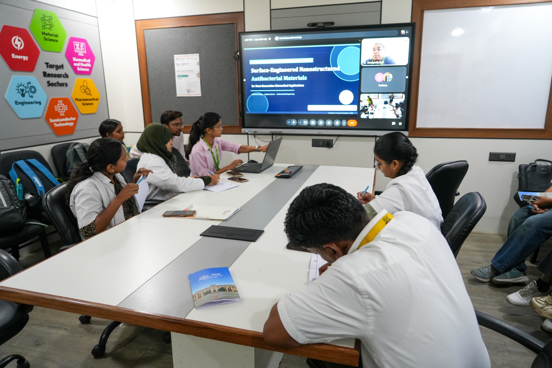
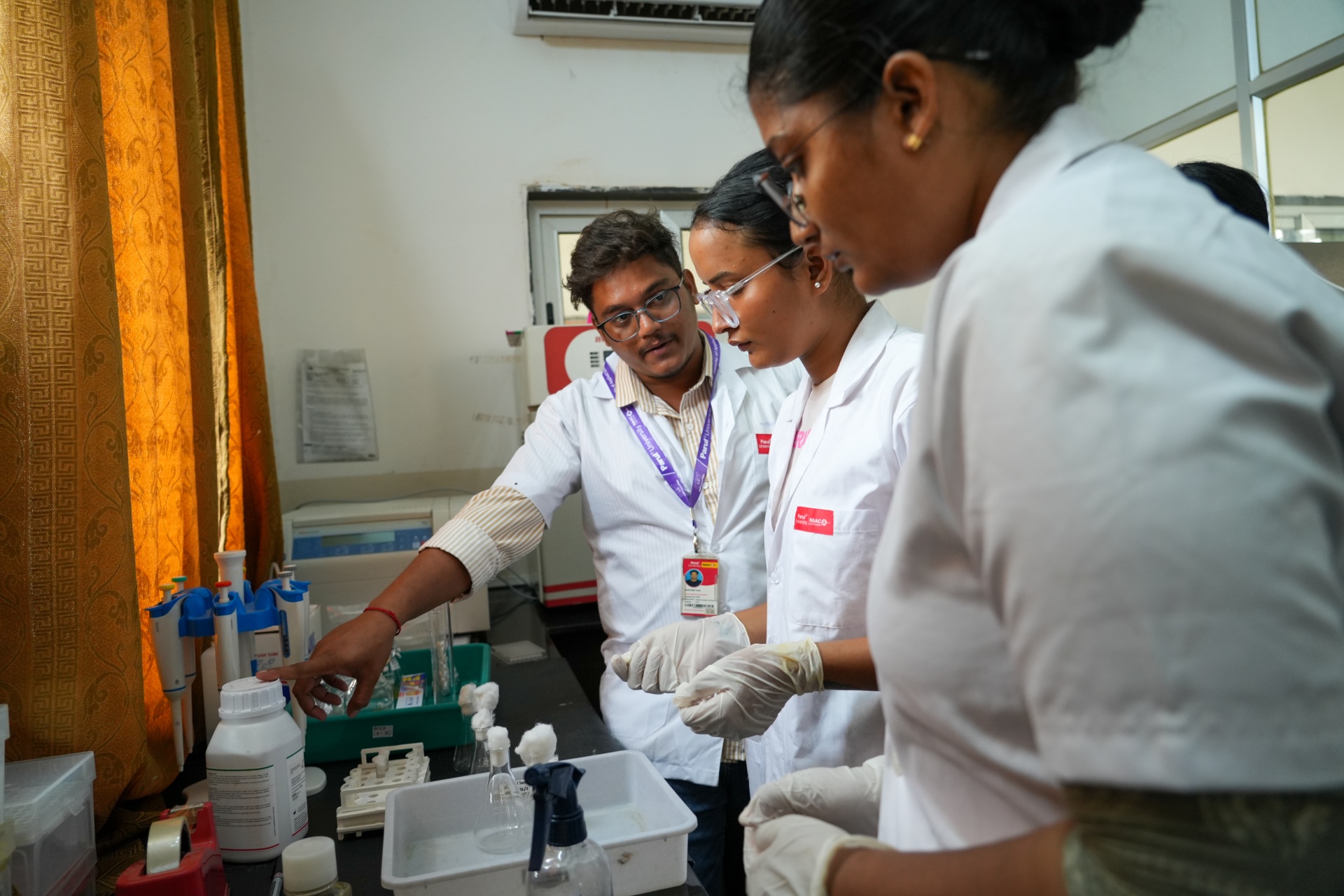
The Microalgae Cultivation track at the MNRDC Three-Day Hands-On Workshop on Microbial Cell Factories ran across 11, 12, and 13 June 2026 at the Micro Nano Research and Development Centre (MNRDC) at Parul University. Dr Anwesha led the track with Mohit Sir delivering the V-770 spectrophotometer operations training on Day 2.
The students got an opportunity to get real time hands-on experience of the complete pipeline from BG-11 media preparation via chlorella via different modes, pigment quantification via Bligh and Dyer lipid extraction. This entire work is positioned across microalgae as biological factories for biofuels, nutraceuticals, anticancer compounds, and high-value biochemicals.
The workshop hub coverage spanning both technical tracks and the two expert sessions is documented at the workshop master hub. The parallel MIC Antimicrobial Assay track is documented at the MIC methodology article. The commercial scale-up context for microalgae cultivation, including Spirulina manufacturing for global markets, is covered in Dr Sanjiv Kumar Mishra’s industry session documented at the Sea Pearl Biotech session coverage. The broader research infrastructure at Parul University including the 7 Stanford-Elsevier top 2 percent scientists is at the faculty currently serving record, with the institutional accreditation framework at the accreditation record.
Day 1: BG-11 media preparation and the foundational cultivation protocol
The very first day was introduced as the BG-11, as their specialised broth is used for growing blue-green algae and freshwater cyanobacteria. Their 11 essential components, BG-11, regularly provide carbon, nitrogen and phosphorus as they’re required for cellular growth and development. Dr Anwesha deliberated that right weighing is critical to measure as deviation usually alters, and so it leads to poor cell growth!
- BG-11 calculation and preparation – The holistic and standard concentration is 1.627 grams of BG-11 P.L., of distilled water. To be precise, 600 ML experimental volume, students calculated an addition of 0.9762 grams of the same powder. The designated flasks for specific nutritional studies, and glucose and sucrose were added as well.
- Carbon source classification – Sugars are classified in two different sets – Monosaccharides and Disaccharides. The standard calculation of heterotrophic media is compulsorily 1 gram of glucose per litre of distilled water, as scaled to 50 ml of volume, and students have successfully measured the expected 0.05 grams of glucose & sucrose.
- Sterilisation methodology. Flask preparation took place inside a Vertical Laminar Airflow Hood forcing HEPA-filtered air in uniform downward direction, creating a sterile environment that prevents contaminated room air settling into open flasks. All working surfaces and hands were cleaned with Ethanol. The Ethanol acts as a rapid disinfectant, denaturing the proteins of any stray microbes present on the gloves or skin. Flask mouths were sealed tightly with dense cotton plugs that allow gas exchange (oxygen and CO2 for algal respiration) while trapping dust and bacteria. Sealed flasks were autoclaved for 1 hour to destroy any latent spores or bacteria present in the distilled water or dry media powder.
3 peak modes - autotrophy, heterotrophy and mixotrophy
Microalgal metabolic flexibility ensures a holistic cultivation across 3 modes, producing different growth ratios and biochemical solutions. This workshop ensured students were bifurcated and they prepared media into flasks and tested 3 modes, they even provided data on how species of Chlorella responds to multi-faceted energy and carbon source conditions!
- The standard growth mode for algae. Subsequently, cells are usually relying on light for major energy and inorganic carbon dioxide from the air. This exclusive mode repeats how algae thrive in natural environments in sync with photosynthesis and metabolic processes.
- Heterotrophy – Usually, cells are thriving in dark mode and provide an organic carbon source. This exclusive workshop covers the end to end testing of glycerol, starch, and glucose as heterotrophic carbon sources. By addition of heterotrophic mode, it produces higher lipid accumulation under controlled conditions but it even requires organic substrate output at all the levels.
- A hybrid approach where algae have access to both organic carbon source and light. Mixotrophic conditions often produce rapid growth and high chlorophyll production. While light is present in mixotrophy, it is not strictly required for survival if the carbon source is rich enough, though light significantly boosts the metabolic rate. The mixotrophic mode typically produces the most rapid biomass accumulation across the three modes.
Comparative results across nutritional treatments showed Mixotrophic Sucrose (MS) produced the highest pigment accumulation at 0.847194 absorbance at 665 nm and 0.833241 at 470 nm. Mixotrophic Glucose (MG) produced 0.1562 at 665 nm and 0.182877 at 470 nm. Heterotrophic Glucose (HG) showed variability across batches from 0.160403 to 0.66169 at 665 nm. The control sample baseline was 0.264606 at 665 nm and 0.308882 at 470 nm. Sucrose under combined light and organic carbon conditions emerged as the most efficient carbon source for the tested Chlorella cultures.
Inoculation, growth tracking, and the microbial growth curve
After media sterilisation and cooling, microalgae were introduced into the prepared flasks. Students had received an opportunity to experiment with micropipette, they added 5 ml of live algal culture to each set of 50 ml media flask, ensuring a 1/10th ratio. Since precision in size matters more than anything, too-sparse starts developing population production in a slow manner while too-dense starting populations rapidly depleting nutrients respectively!
Optical Density (OD) measurement at 685 nanometres tracked culture growth across the 24-hour cycle, with readings taken every 4 hours. As algae multiply, the liquid becomes cloudier with the spectrophotometer measuring the light absorbed by suspended cells. Higher absorbance correlates directly with higher cell number in the flask, supporting quantitative tracking of the growth curve.
- Lag phase. When the 5 ml culture is first introduced to fresh BG-11 media, growth does not begin immediately. Cells are in a resting or adjustment phase, sensing the new environment, assessing nutrient availability, and synthesising the specific enzymes needed for division. Cell count remains flat during this period.
- Log (Exponential) phase. Once adjusted, cells enter a period of rapid exponential division. The mathematics of this phase are aggressive: the microbial population doubles at a constant rate as long as nutrients are plentiful and waste products are low. For Aspergillus (a fungus often studied alongside algae), specialised YP2 media triggers the fastest possible growth during the exponential window.
- Stationary phase. Rapid growth consumes available nutrients. Cells excrete waste products that alter the pH of the media. The rate of new cell division slows to match the rate of cell death. The growth curve flattens. This specific part is divided by physiological stress on the organisms, and is the only harvesting window for lipid extraction!
- Death phase. If left unchecked, the toxic environment and total lack of nutrients cause the death rate to overtake division rate, and the population crashes.
Microscopic observation and morphological analysis using SEM
Scanning Electron Microscope (SEM) analysis allowed students to observe Chlorella cells at high resolution. Chlorella cells measured roughly 3 to 4 micrometres in diameter. Inside the spherical cells, pyrenoids (distinct dense dots within the chloroplasts responsible for carbon fixation and starch synthesis) were clearly visible. Comparative observation of eukaryotic Chlorella (with defined nucleus) versus prokaryotic Cyanobacteria (cyanocystic species) showed the cyanobacteria cells were significantly larger but with much lower overall growth rate and multiplication speed in OD tracking. The observation is important for industrial applications where biomass generation speed often matters more than individual cell size.
Day 2: V-770 spectrophotometer pigment quantification
Day 2 of the microalgae track moved to quantitative pigment analysis using the V-770 spectrophotometer, the JASCO instrument designed to measure optical properties across UV-visible and near-infrared spectrums. Mohit Sir led the operations training. The instrument parameters were calibrated to read wavelengths between 300 nm and 800 nm. Biological targets included chlorophyll A and B (peak absorption around 665 nm spanning 550 to 665 nm) and carotenoids (peak absorption at 470 nm).
- Baseline calibration. Before experimental data collection, students filled a cuvette with pure distilled water as the reference baseline. The baseline ensures the machine reads only the optical density of cellular material, not the water itself.
- Sample preparation for spectrophotometry. Students transferred 2 ml of culture from each experimental flask into Eppendorf tubes labelled as: Controlled (CTRL), Mixotrophic Glucose (MG), Heterotrophic Glucose (HG), Mixotrophic Sucrose (MS), and Heterotrophic Sucrose (HS). The labelling supported tracking across the comparative analysis.
- Pigment significance. Carotenoids carry vibrant orange coloration and operate as powerful antioxidants with documented anticancer properties beyond their role in photosynthesis. Chlorophyll concentrations indicate biomass health and photosynthetic capacity. Both pigments have substantive nutritional and pharmaceutical applications, with their extraction representing one pathway from microbial cell factory to public health solution.
Day 3: Bligh and Dyer lipid extraction and biphasic separation
Day 3 of the microalgae track executed the Bligh and Dyer lipid extraction protocol, the gold standard methodology for total lipid extraction from biological tissues since its development in the late 1950s. The protocol uses a carefully proportioned mixture of polar (methanol) and non-polar (chloroform) solvents to disrupt lipid-protein interactions in cell membranes and solubilise the released lipids.
- Solvent mixture preparation. Methanol (polar) disrupts the complex hydrogen bonds and electrostatic forces binding lipids to structural proteins within algal cell membranes. Chloroform (non-polar) penetrates the cellular matrix to dissolve the newly freed neutral lipids. Students prepared a 15 ml working solution maintaining the critical 2:1 chloroform-to-water/ethanol ratio that defines protocol success.
- Initial extraction. 20 ml of methanol and chloroform mixture introduced to 2 grams of raw biomass. Physical mixing maximises surface area contact between solid cellular material and chemical solvents, ensuring optimal mass transfer of lipids from the intracellular compartment into the solvent phase.
- High-speed centrifugation. Samples loaded into the centrifuge and spun at 6000 RPM for 10 minutes. The mechanical force drives heavier cellular debris (cell wall fragments, denatured proteins, unextracted complex carbohydrates) into a tightly packed pellet at the tube bottom. The clear supernatant above contains dissolved lipids, pigments, and the solvent mixture. The centrifugation was repeated twice for maximum recovery.
- Biphasic separation in separating funnel. Supernatant transferred into a glass separating funnel. To induce phase separation, 20 ml of water and 10 ml of ethanol were added. Water dramatically shifts polarity of the mixture, with ethanol preferentially bonding to water and drawing away from chloroform. The homogeneous liquid crashes into two distinct phases: the heavier non-polar phase (dense dark green chloroform containing lipids and chlorophyll) settles at the bottom, while the lighter aqueous phase (water, ethanol, and water-soluble cellular metabolites) sits above. Students carefully controlled the stopcock at the funnel base to drain the dark green lipid-rich chloroform layer into a clean collection vessel without contamination from the upper aqueous layer.
- Spectrophotometric quantification of extracted pigments. A 2 ml aliquot of the extracted sample was analysed in the V-770 spectrophotometer at 554 nm. The reading produced an optical density of 0.2697. Beer-Lambert law application converted the optical density into exact pigment concentration per millilitre of extract, supporting biochemical profile development for the cultivated strain.
Why the stationary phase matters for lipid harvest
The workshop placed heavy emphasis on the relationship between microbial growth phase and lipid yield. Lipids serve as the chemical precursors for biodiesel (third-generation biofuel feedstock) and contain potent bioactive compounds used in anticancer and antioxidant medical therapies. The critical lesson is that lipid harvest must occur during the stationary phase, not during the log phase.
During the log phase, algae use energy to build proteins and divide. Upon entering the stationary phase under nutrient starvation stress, metabolic priorities shift. Instead of protein synthesis for division, cells stockpile energy as dense lipid droplets inside their cell walls to survive the hostile environment. Harvesting at peak physiological stress maximises lipid yield. To break open the tough cellular walls and release the lipids, chemical solvents including ethanol or methanol disrupt the lipid membranes through chemical stress, separating the raw lipids from structural cellular debris.
THE Sustainability Impact Ratings 2026 context for microalgae research
Parul University holds 7th in India and joint 46th worldwide for SDG 4 (Quality Education) with Quality Education score 81.1 in the Times Higher Education Sustainability Impact Ratings 2026. Hands-on workshops including the MNRDC Microbial Cell Factories track contribute to the teaching outcomes of the SDG 4 methodology weights. The Top 10 in India position for SDG 3 (Good Health and Well-Being) connects directly to the workshop themes around anticancer and antioxidant compound development through microalgae cultivation. The 250-acre campus, 24×7 NABH-accredited Parul Sevashram Hospital, 250+ technology laboratories, MNRDC characterisation infrastructure, and the Rs 58.31 crore in government-funded research projects across 315 funded projects support the broader research environment that workshops like this one operate within.
FAQs
What is BG-11 media and how is it prepared for microalgae cultivation?
BG-11 (Blue Green-11) is a specialised nutritional broth used primarily for growing blue-green algae and freshwater cyanobacteria. The media contains 11 essential components that provide the carbon, nitrogen, and phosphorus required for cellular growth and development. The formulation supports the structural integrity of algal cells and provides the raw materials for photosynthesis and reproduction. The standard preparation concentration is 1.627 grams of powdered BG-11 per litre of distilled water. For experimental volumes, the calculation scales proportionally; the MNRDC workshop prepared 0.9762 grams of BG-11 for 600 ml of distilled water. Accurate weighing is critical because deviations alter osmotic balance and nutrient concentration, leading to poor cell growth or culture death. After dissolving BG-11 powder in distilled water, the prepared media is sterilised through autoclave at high-pressure steam for 1 hour, destroying any latent spores or bacteria present in the distilled water or dry media powder. The sterilised media is then ready for inoculation with microalgae cultures.
What is the difference between autotrophy, heterotrophy, and mixotrophy in microalgae cultivation?
Autotrophy is the standard growth mode where cells rely entirely on light for energy (photosynthesis) and inorganic carbon dioxide from the air as the carbon source. This mode mimics natural outdoor algal growth and produces moderate biomass accumulation. Heterotrophy is the dark-growth mode where cells are provided with an organic carbon source for energy (such as glucose, sucrose, glycerol, or starch) and grown without light. Heterotrophic cultivation often produces higher lipid accumulation under controlled conditions but requires organic substrate input. Mixotrophy is the hybrid mode where algae have access to both organic carbon source and light. Mixotrophic conditions typically produce the most rapid biomass accumulation and high chlorophyll production. While light is present in mixotrophy, it is not strictly required for survival if the carbon source is rich enough, though light significantly boosts metabolic rate. At the MNRDC workshop, comparative testing showed Mixotrophic Sucrose produced the highest pigment accumulation at 0.847194 absorbance at 665 nm, identifying sucrose under combined light and organic carbon conditions as the most efficient carbon source for the tested Chlorella cultures.
What microalgae species were cultivated at the MNRDC workshop and what are their industrial applications?
The MNRDC workshop focused on two Chlorella species: Chlorella vulgaris and Chlorella minutissima. Both are highly valued in industrial biotechnology with overlapping and distinct application profiles. Chlorella vulgaris is particularly noted for pigment production with applications involving Multi-Walled Carbon Nanotubes (MWCNT) for advanced material research, alongside its use in nutraceutical and food supplement applications. Chlorella minutissima offers exceptional candidacy for lipid production, with the cell biology supporting accumulation of triacylglycerols (TAGs) as energy reserves under nutrient stress, making the species relevant for biofuel feedstock and high-value bioactive compound production. Beyond Chlorella, the broader microalgae industry uses Haematococcus (source for astaxanthin pigment), Dunaliella salina (source for beta-carotene), Spirulina platensis (source for protein and phycocyanin), Nannochloropsis (source for omega-3 fatty acids including DHA), and Aurantiochytrium, Porphyridium, and Porphyra species for various commercial applications. The commercial scale-up context for microalgae cultivation is covered in the industry expert session by Dr Sanjiv Kumar Mishra (Sea Pearl Biotech) documented separately.